 Apollo Hospitals, Opp.iim 154/11,bannerghatta Road.
 Posted on July 20, 2026
Complex Multivessel Coronary Intervention: A Heart-Team Approach to Calcified Triple-Vessel Disease
Complex coronary artery disease is rarely solved by a single balloon or a single stent. Some patients present with several narrowed vessels, previous coronary implants, hard calcified plaque and medical conditions that increase both procedural and recovery risk. In these situations, the quality of the outcome depends on planning, technology and teamwork as much as it depends on the final angiographic image.
This anonymised case from the Centre of Excellence for Complex Coronary Interventions at Apollo Hospitals, Bengaluru, describes the evaluation and treatment of an older adult with unstable angina, previous angioplasty, diabetes, hypertension and triple-vessel coronary artery disease. The intervention combined orbital atherectomy, sequential balloon preparation, overlapping drug-eluting stents in the right coronary artery and drug-coated balloon therapy in the left circumflex artery.
The case is especially valuable because it demonstrates how a multidisciplinary team can individualise treatment when different vessels require different solutions. Rather than treating every narrowing in the same way, the strategy was adapted to the plaque characteristics, previous stents, vessel size, bifurcation anatomy and overall bleeding risk.
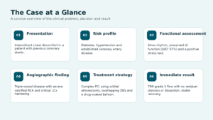
Figure 1. The anonymised clinical case at a glance.
1. Clinical Background: Why New Chest Discomfort Needed Reassessment
The patient had established coronary artery disease and had undergone angioplasty with stents to the proximal-to-mid left anterior descending artery and the left circumflex-to-obtuse marginal segment several years earlier. The previous procedure had provided durable benefit, but a history of coronary intervention does not prevent new plaque from developing elsewhere or within the margins of treated segments.
The patient later reported intermittent chest discomfort. The symptoms were not continuously severe, yet they were clinically important because of the previous coronary history and the presence of major risk factors. Diabetes can accelerate atherosclerosis and may also produce less typical warning symptoms. Hypertension adds further stress to the vascular system and increases long-term cardiovascular risk.
An electrocardiogram showed sinus rhythm. Echocardiography demonstrated preserved left ventricular systolic function, with an ejection fraction of approximately 57%. Preserved pumping function was reassuring, but it did not exclude important coronary narrowing. A stress test was positive, indicating inducible myocardial ischaemia and supporting the need for coronary angiographic evaluation.
|
Important clinical principle A normal or preserved ejection fraction does not mean the coronary arteries are normal. A patient can have significant flow-limiting disease while the heart’s resting pumping function remains preserved. |
2. Angiography Revealed Triple-Vessel Coronary Disease
Coronary angiography demonstrated disease across all three major coronary territories. The previously implanted stents remained largely patent, but new and progressive narrowing was present beyond and around the treated segments.
Left anterior descending artery
The proximal-to-mid LAD stent was open with mild in-stent restenosis. Beyond the stented segment, a long 70% to 80% tubular narrowing was seen. The ramus intermedius also showed moderate proximal and mid-vessel disease.
Left circumflex artery
The left circumflex was non-dominant but had a severe ostial-to-proximal narrowing of approximately 90%. The earlier LCx-to-obtuse-marginal stent remained patent. Distally, the circumflex was a smaller vessel with an additional moderate narrowing.
Right coronary artery
The dominant right coronary artery contained diffuse disease: approximately 80% narrowing proximally, 70% in the mid segment and a further 60% lesion distally. The plaque was heavily calcified, which substantially increased the technical difficulty of balloon expansion and stent delivery.
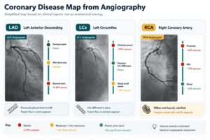
Figure 2. Simplified disease map based on the coronary angiogram. This is a schematic and not a patient-specific anatomical tracing.
3. Heart-Team Decision: Medical Therapy or Complex PCI?
The angiographic findings were reviewed together with the patient’s symptoms, positive stress test, preserved ventricular function, previous interventions and medical comorbidities. The team discussed the implications of the disease and the available management pathways, including optimised medical therapy and percutaneous coronary intervention.
The decision was not based only on the percentage of narrowing. The team considered symptom burden, evidence of ischaemia, vessel dominance, lesion length, calcification, prior stent architecture, the need for prolonged antiplatelet therapy and the likelihood of achieving a safe and durable result.
After a detailed discussion of expected benefits, limitations and risks, the patient and family chose complex angioplasty. The plan prioritised treatment of the dominant calcified right coronary artery and the severe circumflex lesion while preserving previously treated segments and avoiding unnecessary additional metal where a drug-coated balloon could be used.
|
Why “one plan for every vessel” would not be appropriate The RCA required calcium modification and scaffolding with stents. The LCx required a different approach because of its location, prior stent relationship and bifurcation considerations. Tailoring the device strategy to each vessel is a defining feature of complex PCI. |
4. Why This Procedure Was Technically Demanding
Several features placed this intervention in the complex PCI category:
- Diffuse multivessel disease rather than one short, focal blockage.
- Severe calcification in the dominant right coronary artery.
- Multiple sequential lesions extending from the proximal to distal RCA.
- Previous LAD and LCx-to-OM stents that had to be protected and incorporated into the new strategy.
- A severe ostial/proximal LCx lesion near a bifurcation, where treatment of one branch can influence the other.
- Diabetes and hypertension, which increase long-term cardiovascular risk.
- A low platelet count that required haematology input to balance bleeding risk against the need for antiplatelet therapy.
The combination of calcium, vessel tortuosity, long lesion length and previous implants meant that device delivery and complete stent expansion could not be taken for granted. The team therefore planned stepwise lesion preparation and used guide-extension and microcatheter support where required.
5. The Procedure: Step-by-Step
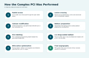
Figure 3. Procedural sequence used during the complex multivessel intervention.
Radial access and guide support
The intervention was performed through the right radial artery using a 7F sheath. Radial access can improve patient comfort and may reduce access-site bleeding in appropriately selected patients, but complex procedures still require careful guide-catheter selection and adequate support.
A workhorse coronary wire was used to cross the lesion. A dedicated atherectomy wire was then positioned with the help of a microcatheter. This sequence provided controlled access across the calcified segment while maintaining the support needed for subsequent devices.
Orbital atherectomy for calcium modification
The right coronary plaque was too rigid for routine balloon treatment alone. Orbital atherectomy was therefore performed at 80,000 revolutions per minute in four controlled runs of approximately 25 seconds each. The objective was not to remove the entire plaque. Instead, the rotating crown modified superficial calcium and increased vessel compliance so that balloons and stents could expand more effectively.
Sequential balloon preparation
Following atherectomy, the RCA was prepared progressively using small-diameter balloons, followed by a cutting balloon. A guide-extension catheter was used to improve device support. Gradual preparation reduced the risk of forcing a large balloon or stent through an inadequately modified lesion.
Overlapping drug-eluting stents in the RCA
Two drug-eluting stents were implanted to cover the long diseased segment from the proximal through the distal RCA. A 3.5 × 26 mm stent was positioned proximally and overlapped with a 3.0 × 38 mm stent extending into the mid-to-distal vessel. Non-compliant balloon post-dilatation was then performed to optimise expansion and apposition.
Drug-coated balloon treatment of the LCx
The left circumflex lesion was crossed and pre-dilated with a 2.75 mm balloon. A 3.0 × 25 mm drug-coated balloon was then inflated for approximately 90 seconds. This delivered an antiproliferative medication to the vessel wall without leaving an additional permanent stent layer in the treated segment.
Bifurcation optimisation and final angiography
Because the LCx lesion was close to the LAD–LCx bifurcation and previous stents, the team performed kissing-balloon optimisation. Targeted post-dilatation was performed in both branches to preserve geometry and flow. The final angiogram showed TIMI grade 3 distal flow with no significant residual stenosis and no angiographic dissection.
6. Right Coronary Artery: Treating Diffuse Calcified Disease
The dominant RCA supplied a large myocardial territory and contained several sequential stenoses. The before-and-after angiographic views illustrate the difference between an artery with diffuse calcified narrowing and the final result after lesion preparation, stenting and optimisation.
A central challenge in calcified PCI is preventing stent under-expansion. A stent that appears to be in the correct position may still perform poorly if rigid calcium prevents it from opening symmetrically. The team addressed that risk before implantation through atherectomy, sequential ballooning and cutting-balloon preparation, rather than relying on high-pressure expansion alone.
The final RCA result demonstrated restored lumen continuity and brisk distal contrast flow. Equally important, the report documented no residual stenosis or dissection, supporting a controlled and complete immediate result.
7. Left Circumflex Artery: Treating a Severe Lesion Without Another Stent Layer
The circumflex lesion presented a different problem. It was severe and located near the vessel origin, with a previously treated LCx-to-OM segment nearby. Adding another metal layer can sometimes complicate future access, alter bifurcation geometry or create a long multilayered segment. The team therefore selected a drug-coated balloon after adequate lesion preparation.
Figure 5. Representative LCx angiography before and after balloon preparation, drug-coated balloon treatment and bifurcation optimisation.
A drug-coated balloon is not simply a standard balloon used at a higher pressure. The lesion must first be prepared so the balloon can contact the vessel wall effectively. The medicated balloon is then inflated for a sustained period to transfer an antiproliferative drug. After deflation, the balloon is removed and no permanent scaffold remains.
This approach was combined with kissing-balloon optimisation to protect the adjoining branch and improve the final bifurcation result. The post-treatment angiogram showed improved lumen and distal flow without angiographic dissection.
8. Why Three Advanced Technologies Were Combined
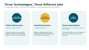
Figure 6. Each technology addressed a different component of the coronary disease.
Orbital atherectomy: making rigid calcium more treatable
Orbital atherectomy was used for lesion preparation. Its purpose was to modify calcium and create a more compliant pathway for balloons and stents. It did not replace angioplasty; it made the subsequent angioplasty more predictable.
Drug-eluting stents: supporting a long diseased segment
The long RCA lesion required continuous scaffolding. Drug-eluting stents restored the vessel lumen, sealed the treated plaque and released medication designed to reduce excessive tissue regrowth. Overlap was necessary to avoid leaving a gap between the two treated zones.
Drug-coated balloon: local therapy without permanent metal
The LCx was treated with a drug-coated balloon so that medication could be delivered without placing another stent layer. This strategy is particularly useful only when lesion preparation is satisfactory and the final vessel result is stable, with acceptable residual narrowing and no major dissection.
9. The Centre of Excellence Team Behind the Procedure
Although the angioplasty is performed in the catheterisation laboratory, the clinical outcome reflects the work of a wider system. This case required coordination before, during and after the intervention.
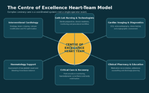
Figure 7. The multidisciplinary heart-team model supporting complex coronary intervention at Apollo Hospitals, Bengaluru.
Interventional cardiology
The interventional team reviewed the angiogram, selected the target vessels, planned wire and guide support, chose the calcium-modification technique, determined stent lengths and diameters and decided where a drug-coated balloon was preferable to another stent.
Cath-lab nursing and technologists
Complex PCI depends on device preparation, sterile workflow, anticoagulation checks, haemodynamic monitoring and rapid availability of balloons, wires, microcatheters, guide extensions, atherectomy equipment and emergency support. Cath-lab nurses and technologists maintain this procedural coordination throughout the case.
Diagnostic and imaging support
ECG, echocardiography, stress testing and angiographic interpretation established the clinical context. These investigations helped the team separate preserved heart-pumping function from the presence of important flow-limiting coronary disease.
Haematology collaboration
A low platelet count required specialist review. This was relevant because stent treatment requires antiplatelet therapy, while thrombocytopenia may increase bleeding risk. Haematology input helped the team balance these competing concerns and define safe monitoring and follow-up.
Recovery, pharmacy and patient education
After the procedure, the patient required access-site monitoring, assessment for recurrent symptoms, review of kidney function and blood counts, glucose and blood-pressure control, medication reconciliation and counselling about the importance of uninterrupted cardiac therapy. These steps are essential to protect the procedural result after discharge.
|
What “Centre of Excellence” should mean in practice It is not only a label for performing advanced procedures. It should represent structured case review, access to specialised technology, experienced cath-lab support, collaboration across departments, standardised recovery pathways and clear long-term follow-up. |
10. Hospital Course and Immediate Outcome
The patient remained haemodynamically stable after the procedure. The radial access site was monitored, medications were optimised and the haematology recommendations were followed. No procedural complication was reported during the remaining hospital stay.
The patient was ambulatory at discharge with stable vital signs, controlled discomfort and a clean access site. Cardiac medications, diabetes treatment, blood-pressure therapy and intensive lipid-lowering treatment were continued or adjusted according to the treating team’s plan.
The immediate technical endpoint was strong: TIMI grade 3 distal flow in the treated vessels, without significant residual stenosis or angiographic dissection. However, the team also emphasised that a technically successful procedure must be followed by disciplined medication adherence and risk-factor management.
Immediate post-procedure status
|
Stable haemodynamics Monitored without instability |
TIMI grade 3 flow Brisk distal perfusion |
No residual stenosis Final lumen satisfactory |
|
No angiographic dissection Vessel integrity preserved |
Ambulatory at discharge Early mobilisation achieved |
Radial access site clean No reported access complication |
11. Recovery, Medication Adherence and Warning Signs
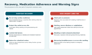
Figure 8. Core recovery priorities and symptoms that require urgent medical review.
Following complex PCI, the most important instruction is not to stop prescribed cardiac medicines without direct advice from the treating cardiology team. Dual antiplatelet therapy protects newly implanted stents from clot formation. Even a brief unplanned interruption may carry risk, particularly in the early period after intervention.
Patients are generally advised to avoid heavy lifting, pulling, pushing and straining while the radial access site heals. Exercise should be restarted gradually and only as advised. A low-salt, low-saturated-fat, high-fibre diet, together with diabetes, blood-pressure and cholesterol control, supports long-term cardiovascular health.
Urgent evaluation is necessary for recurrent chest pain, sweating, shortness of breath, palpitations, marked dizziness, fainting or severe uneasiness. Because blood-thinning medicines are required, bleeding from any site, dark urine, black stools, blood in vomit or sputum, or a severe unexplained headache should also be reported promptly.
12. What Patients and Families Can Learn from This Case
- Previous stents can remain open while new disease develops elsewhere. Long-term follow-up remains important even after a successful earlier angioplasty.
- Preserved heart function does not exclude severe coronary disease. Symptoms and stress-test findings must be evaluated in context.
- Calcified coronary blockages may require specialised lesion preparation before a stent can be implanted safely and expanded fully.
- Not every blockage requires the same device. A stent may be appropriate in one vessel while a drug-coated balloon is selected for another.
- Complex PCI is safest when performed within a coordinated system that includes interventional expertise, cath-lab technology, nursing support, diagnostics, critical care and access to other specialties.
- The long-term result depends on more than the procedure. Medication adherence, diabetes and blood-pressure control, lipid lowering, diet, activity and follow-up all remain essential.
13. Frequently Asked Questions
Is triple-vessel disease always treated with bypass surgery?
No. Some patients are best treated with coronary bypass surgery, some with medication and some with PCI. The decision depends on coronary anatomy, diabetes status, ventricular function, symptom severity, surgical risk, lesion complexity, patient preference and the likelihood of achieving complete and durable revascularisation. A heart-team review is particularly valuable when more than one strategy is reasonable.
Why was atherectomy needed before the RCA stents?
The RCA plaque was heavily calcified. Rigid calcium can prevent balloons and stents from expanding properly. Orbital atherectomy modified the calcified surface and improved vessel compliance before ballooning and stent implantation.
Why were two stents used in the RCA?
The disease extended across a long segment from the proximal through the distal RCA. One stent would not have covered the entire diseased zone. Two appropriately sized stents were overlapped to avoid leaving an untreated gap.
Why was the LCx treated with a drug-coated balloon instead of another stent?
The LCx lesion was near a previously treated segment and a bifurcation. After satisfactory lesion preparation, a drug-coated balloon allowed medication to be delivered without adding another permanent metal layer. This decision is anatomy-specific and cannot be applied to every coronary narrowing.
What does TIMI grade 3 flow mean?
TIMI grade 3 describes normal or near-normal forward blood flow through the treated artery on angiography. It is an important immediate procedural endpoint, although long-term success still depends on healing, medication adherence and control of cardiovascular risk factors.
Can coronary disease return after a successful procedure?
Yes. The treated segment can develop restenosis, and new plaque can develop in other parts of the coronary circulation. Angioplasty treats specific blockages; it does not remove the underlying tendency to atherosclerosis. Long-term prevention therefore remains essential.
Conclusion: Advanced Technology Works Best Within an Experienced Team
This anonymised case demonstrates how complex multivessel coronary disease can require several complementary techniques within the same procedure. Orbital atherectomy prepared a rigid, calcified RCA. Sequential balloons and a cutting balloon created a safer foundation for two overlapping drug-eluting stents. A severe LCx lesion was treated using a drug-coated balloon and bifurcation optimisation, avoiding an unnecessary additional stent layer. Final angiography showed TIMI grade 3 flow without residual stenosis or dissection.
The broader message is equally important: complex coronary intervention is not defined only by the devices used. It is defined by appropriate patient selection, careful angiographic planning, skilled execution, multidisciplinary collaboration and structured follow-up.
At the Centre of Excellence for Complex Coronary Interventions, Apollo Hospitals, Bengaluru, complex cases are evaluated through a team-based pathway designed to identify the safest and most effective treatment strategy for each patient’s anatomy and overall medical condition.
For patients seeking an evaluation
Patients with recurrent chest discomfort, previous stents, diabetes, complex coronary disease or a recommendation for advanced angioplasty may benefit from a detailed heart-team assessment. Treatment decisions must be made after personal clinical examination and review of the original ECG, echocardiogram, stress test and coronary imaging.