What is Optical Coherence Tomography (OCT) Imaging?
Optical Coherence Tomography (OCT) is an intravascular imaging modality that uses near-infrared light to provide high-definition, cross-sectional and three-dimensional images of the vessel microstructure during angioplasty.
“The images provide additional information on the degree and characteristics of coronary artery disease compared to angiography which doesn’t delineate the composition of the coronary artery. With automated, highly accurate measurements, OCT can guide stent selection, placement, and deployment.”
How does OCT support angioplasty guidance?
- OCT can be used to identify the content and size of block precisely to select right balloon and stent.
- For post angioplasty guidance, OCT helps to confirm that stent is fully expanded to reduce stent failure.
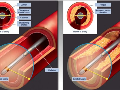
IVUS:
Intravascular ultrasound is an intravascular imaging modality primarily used in interventional cardiology to characterize lesion morphology, quantify plaque burden, guide stent sizing, assess stent expansion, and identify procedural complications. Using a dedicated catheter with ultrasound-based technology an image is obtainable from inside an artery producing cross-sectional images with a 360-degree view of the vessel. This technology overcomes many of the limitations of angiography, which utilizes x-ray technology to produce a 2-dimensional lumenogram of a 3-dimensional structure. Rather than assessing the vessel from the outside as with angiography, IVUS provides the assessment from within the vessel.