 Posted on July 22, 2024
Understanding Angiograms: Femoral, Radial, and Ulnar Approaches
Introduction
Angiograms, also known as angiographies, are medical imaging techniques used to visualize the inside of blood vessels and organs, particularly the arteries, veins, and heart chambers. This procedure helps diagnose and treat various cardiovascular conditions by providing detailed images of the blood flow and identifying any blockages or abnormalities. There are three primary approaches to performing an angiogram: femoral, radial, and ulnar. Each approach has its advantages and specific uses, and understanding them can help patients and healthcare providers make informed decisions.
Femoral Approach
Procedure
The femoral approach involves accessing the femoral artery, which is located in the groin area. Here’s how the procedure typically unfolds:
- Preparation: The patient is positioned on the examination table, and the groin area is sterilized and numbed with a local anesthetic.
- Insertion: A small incision is made, and a catheter is inserted into the femoral artery.
- Navigation: Using fluoroscopy (real-time X-ray imaging), the catheter is guided through the arterial system to the area of interest.
- Imaging: A contrast dye is injected through the catheter, and X-ray images are taken to visualize blood flow and identify any blockages or abnormalities.
Advantages
- Direct Access: The femoral artery provides a large and direct access point to the heart and major blood vessels, making it ideal for complex procedures.
- Familiarity: It is a well-established approach with a long history of use in angiography and interventional cardiology.
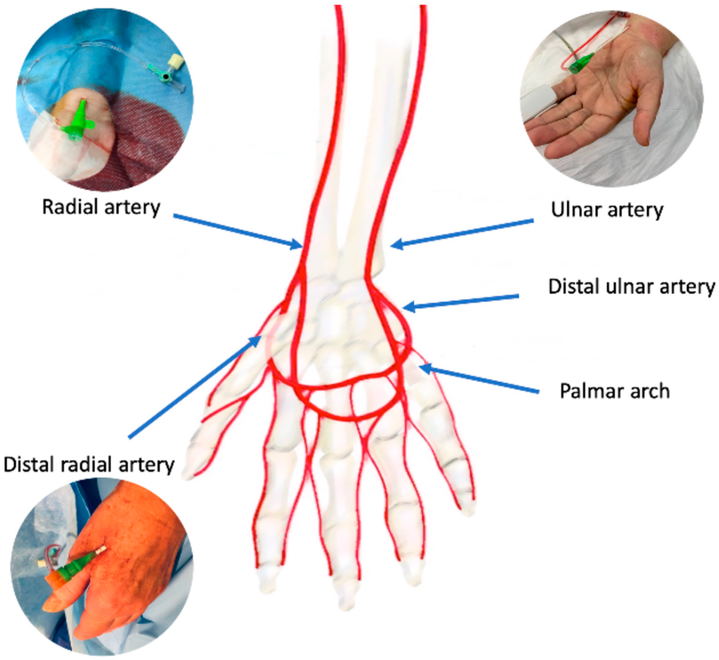
Radial Approach
Procedure
The radial approach involves accessing the radial artery in the wrist. The steps are as follows:
- Preparation: The patient’s arm is positioned and sterilized, and a local anesthetic is administered to the wrist area.
- Insertion: A small needle is used to puncture the radial artery, and a catheter is inserted.
- Navigation: The catheter is guided through the radial artery to the coronary arteries or other areas of interest.
- Imaging: Similar to the femoral approach, a contrast dye is injected, and X-ray images are taken.
Advantages
- Comfort and Mobility: Patients can sit up and move around soon after the procedure, enhancing comfort and reducing the need for prolonged bed rest.
- Lower Complication Rates: There is a reduced risk of bleeding and vascular complications compared to the femoral approach.
Ulnar Approach
Procedure
The ulnar approach is similar to the radial approach but involves accessing the ulnar artery, also located in the wrist but on the opposite side of the radial artery. The procedure steps include:
- Preparation: The ulnar artery site is sterilized, and a local anesthetic is applied.
- Insertion: A catheter is inserted into the ulnar artery.
- Navigation: The catheter is guided to the area of interest using fluoroscopic guidance.
- Imaging: A contrast dye is injected, and X-ray images are obtained.
Advantages
- Alternative Access: It provides an alternative access site if the radial artery is not suitable or has been previously used extensively.
- Patient Comfort: Like the radial approach, it allows for quicker mobilization post-procedure.
Choosing the Right Approach
The choice between femoral, radial, and ulnar approaches depends on various factors, including:
- Patient Anatomy: Some patients may have anatomical variations that make one approach more suitable than the others.
- Procedure Complexity: Complex interventions may benefit from the femoral approach due to its direct and larger access point.
- Patient Preference: Patient comfort and recovery preferences play a significant role in the decision-making process.
- Physician Expertise: The experience and training of the healthcare provider in a specific approach can influence the choice.