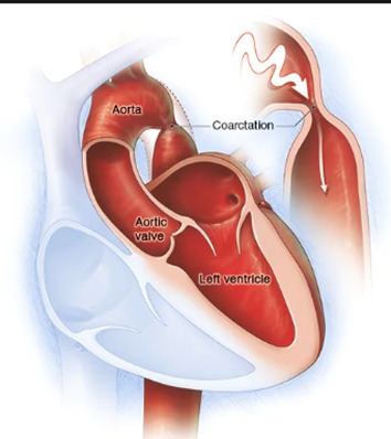
AORTIC COARCTATION
The aorta is the largest artery in the body. It moves oxygen-rich blood from the heart to the rest of the body. Aortic coarctation is a narrowing of the aorta. It forces the heart to pump harder to move blood through the aorta.
Coarctation of the aorta is generally present at birth (congenital heart defect). Symptoms can range from mild to severe. The condition might not be detected until adulthood.
Coarctation of the aorta often occurs along with other congenital heart defects. Treatment is usually successful, but the condition requires careful lifelong follow-up.
Causes
The cause of coarctation of the aorta is unclear. The condition is generally a heart problem present at birth (congenital heart defect).
Rarely, coarctation of the aorta develops later in life. Conditions or events that can narrow the aorta and cause this condition include:
- Traumatic injury
- Severe hardening of the arteries (atherosclerosis)
- Inflamed arteries (Takayasu arteritis)
Coarctation of the aorta can affect any part of the aorta, but it’s most often located near a blood vessel called the ductus arteriosus. That blood vessel connects the left pulmonary artery to the aorta.
With coarctation of the aorta, the left lower heart chamber (left ventricle) works harder to pump blood through the narrowed aorta. As a result, blood pressure rises in the left ventricle. The wall of the left ventricle may become thick (hypertrophy).
There are several procedures and surgeries to repair aortic coarctation. Together, you and your health care team can discuss which type is most likely to be successful. Options include:
Balloon angioplasty and stenting. This may be the first treatment for aortic coarctation. Sometimes it’s done if narrowing occurs again after coarctation surgery.
During balloon angioplasty, the doctor inserts a thin, flexible tube (catheter) into an artery in the groin. It’s moved through the blood vessels to the heart using X-rays as a guide. An uninflated balloon is placed into the catheter and moved into the area of the narrowed aorta. The balloon is inflated. The inflated balloon widens the aorta, so blood flows more easily. Angioplasty is often combined with the placement of a small wire mesh tube called a stent. The stent helps keep the artery open, decreasing the chance of narrowing again.
Resection with end-to-end anastomosis. This method involves removing the narrowed area of the aorta (resection) and then connecting the two healthy parts of the aorta (anastomosis).
Subclavian flap aortoplasty. A part of the blood vessel that delivers blood to the left arm (left subclavian artery) might be used to expand the narrowed area of the aorta.
Bypass graft repair. This surgery uses a tube called a graft to reroute blood around the narrowed area of the aorta.
Patch aortoplasty. The surgeon cuts across the narrowed area of the aorta and then attaches a patch of synthetic material to widen the blood vessel. Patch aortoplasty is useful if the coarctation involves a long part of the aorta.