 Apollo Hospitals, Opp.iim 154/11,bannerghatta Road. Bannerghatta Road.bangalore-560076
 6366 229563
 Posted on August 31, 2026
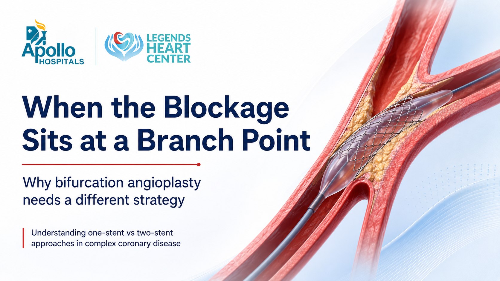
When the Blockage Sits at a Branch Point: Why This Angioplasty Needs a Different Strategy
A patient may hear after an angiogram:
“The blockage is not only severe — it is sitting exactly where the artery divides.”
That single detail can completely change how angioplasty is planned.
A blockage at a coronary artery branch point is called a bifurcation lesion. It is more challenging than a straightforward narrowing because the cardiologist is not trying to protect just one blood vessel. Two important pathways may need to remain open at the same time.
The encouraging part is that many bifurcation blockages can still be treated successfully using modern stenting techniques, advanced imaging and careful procedural planning.
The real question is usually not:
“Can a stent be placed?”
It is:
“How do we treat the main blockage without compromising the important side branch?”
Think of It Like a Road Junction
Imagine a major road that reaches a Y-junction.
One road continues straight ahead. Another important road branches away from it.
Now imagine a large obstruction developing exactly at that junction.
Clearing only the main road could push material toward the second road and partially close it. Trying to widen both roads without a proper plan could make the junction unstable.
The same principle applies inside the coronary arteries.
A bifurcation has a main vessel and a side branch, and both may supply important areas of heart muscle.
That is why location matters so much.
What Makes a Branch-Point Blockage Different?
In a simple coronary narrowing, the cardiologist may be able to treat one segment with a balloon and stent.
At a bifurcation, the procedure has another important consideration: what happens to the side branch after the main vessel is treated?
When a stent expands, plaque may shift. The opening of the side branch may narrow. The angle between the vessels may change.
For some patients, that side branch is relatively small and losing a little blood flow may not be clinically important.
For others, the branch supplies a large area of heart muscle and protecting it becomes a major priority.
This is why two angiograms that both say “80% blockage” may require very different procedures.
The First Big Decision: One Stent or Two?
This is one of the most important parts of bifurcation angioplasty.
Patients sometimes assume that treating two branches automatically requires two stents.
Often, it does not.
When One Stent May Be Enough
For many bifurcation lesions, cardiologists prefer a provisional stenting strategy.
The main vessel is treated first while access to the side branch is preserved with a guidewire.
After the main stent is placed, the side branch is reassessed.
If blood flow remains good and the branch opening is satisfactory, no second stent may be required.
If the branch becomes significantly narrowed or compromised, the cardiologist can then treat it.
This approach allows the procedure to remain as simple as possible while keeping a second-stent option available.
When Two Stents May Be Planned From the Beginning
Some bifurcations are genuinely complex.
If both branches have substantial disease, the side branch is large, or the narrowing extends well into both vessels, treating only the main artery may not produce an adequate result.
In that situation, the cardiologist may decide that both branches need stents.
Techniques may include approaches known as:
DK Crush, Culotte, T-stenting or TAP.
The names sound technical, but the underlying goal is straightforward:
keep both important branches open while creating a stable, well-expanded stent result.
The technique selected depends on the anatomy rather than personal preference alone.
Why “Just Put Another Stent” Is Not Always the Best Answer
In bifurcation PCI, more metal inside the artery is not necessarily better.
Every additional stent adds another layer of complexity.
Two-stent procedures require precise positioning and careful optimisation. Poor expansion or overlapping stents in the wrong position can increase the risk of restenosis or other stent-related problems.
That is why experienced operators generally try to use the simplest strategy capable of producing a safe and durable result.
Sometimes that means one stent.
Sometimes it means two.
What matters is choosing correctly.
A Small Technical Detail Can Make a Big Difference: POT
One phrase patients may hear during complex PCI is POT — Proximal Optimization Technique.
The artery before a branch point is usually larger than the artery after the branch.
A stent sized for the smaller vessel may therefore not fully touch the vessel wall in the larger proximal section.
POT uses a carefully selected balloon to optimise that part of the stent.
Although it sounds like a small technical step, it can help improve stent expansion and make it easier to maintain access to the side branch.
In complex bifurcation angioplasty, details such as these can strongly influence the final result.
What Is “Kissing Balloon” Inflation?
Another term that can sound unusual is kissing balloon inflation.
Two balloons are positioned — one in each branch — and inflated in a coordinated way.
This can help optimise the opening of both branches after certain bifurcation stenting procedures.
It is not required in every case, but it may form an important part of selected one- or two-stent strategies.
Why IVUS or OCT Can Be Especially Valuable Here
An angiogram shows the outline of the coronary artery.
But in a complicated branch point, the cardiologist may need more information than an X-ray image alone can provide.
IVUS
Intravascular Ultrasound uses a miniature ultrasound probe inside the artery.
It can help assess the true vessel size, calcium, plaque distribution, stent expansion and whether the stent is sitting correctly.
It can be particularly useful in complex disease and left-main interventions.
OCT
Optical Coherence Tomography provides highly detailed images from inside the artery.
It may help the cardiologist evaluate stent apposition, under-expansion, edge problems and how stent struts interact with the side branch.
In complex bifurcation PCI, imaging is often less about “adding technology” and more about reducing uncertainty.
What If the Branch Point Is in the Left Main Artery?
This deserves special attention.
The left main coronary artery supplies a large portion of the heart.
It eventually divides into two major vessels: the left anterior descending artery and the circumflex artery.
When a blockage sits at this division, the consequences of treatment are potentially more significant because both branches are important.
Some patients may be better treated with bypass surgery.
Others, particularly selected patients with suitable anatomy or increased surgical risk, may be considered for left-main PCI.
The decision may depend on coronary anatomy, diabetes, heart function, other blockages, age, surgical risk and the patient’s overall condition.
This is one situation where an experienced Heart Team discussion can be particularly useful.
What If Calcium Is Also Present?
Severe calcium can make bifurcation angioplasty substantially more difficult.
A heavily calcified artery may not expand properly even when a balloon is inflated.
If the vessel is not prepared adequately, the stent may remain under-expanded.
Depending on the anatomy, calcium-modification techniques such as intravascular lithotripsy, specialised balloons or atherectomy may be considered before stenting.
The more complicated the lesion, the more important preparation becomes.
A Patient Scenario
Consider a patient who has significant disease where a large coronary artery divides into two branches.
The written angiogram report may simply say:
“Severe bifurcation stenosis.”
But the cardiologist still needs to answer several questions.
Is the side branch large enough to protect aggressively?
How long is the blockage in the branch?
Is there calcium?
Is one stent likely to preserve the second branch?
Would a planned two-stent strategy be safer?
Would IVUS help size the vessel?
Would bypass surgery provide a better long-term result?
The procedure begins long before the first balloon enters the artery.
It begins with understanding the anatomy.
The Goal Is Not to Put in More Stents
This is worth emphasizing.
Successful complex angioplasty is not judged by how many stents are used.
The aim is to achieve:
good blood flow, adequate stent expansion, preservation of important branches and a durable result with the least unnecessary complexity.
For patients, this is the most useful way to think about bifurcation PCI.
Frequently Asked Questions
1. Is a bifurcation blockage more dangerous than a normal blockage?
Not automatically. It is usually more technically complex to treat because two branches may be involved. The clinical importance depends on the size of the vessels, severity of the narrowing and how much heart muscle they supply.
2. Does a branch-point blockage always require two stents?
No. Many bifurcation lesions can be treated using a one-stent provisional strategy. A second stent is added only when the side branch requires treatment.
3. Why would a cardiologist avoid placing two stents immediately?
Using two stents makes the procedure more complex and introduces more overlapping metal inside the artery. When one stent can provide an excellent result, adding another may not offer additional benefit.
4. Can the side branch close during angioplasty?
It can become narrowed or occasionally lose blood flow after main-vessel stenting. Cardiologists use guidewires, balloon techniques, imaging and specific bifurcation strategies to reduce and manage this risk.
5. What determines whether the side branch is important?
Its size, the amount of heart muscle it supplies and the severity and length of disease inside it are all important considerations.
6. What is a “true bifurcation lesion”?
This generally refers to disease involving both the main vessel and the important side branch around the point where they divide. These lesions can require more complex planning.
7. Is left-main bifurcation angioplasty possible?
Yes, in selected patients. However, left-main bifurcation disease is particularly important and treatment may involve either PCI or bypass surgery depending on anatomy and overall clinical factors.
8. Does IVUS make bifurcation angioplasty safer?
IVUS can provide valuable information about vessel size, calcium and stent expansion. It is especially useful in many complex and left-main procedures, although the need for imaging is determined individually.
9. What happens if the blockage is also heavily calcified?
The artery may need to be prepared before stenting using calcium-modification techniques. This helps the stent expand more effectively.
10. Is bifurcation angioplasty more likely to fail?
Complex bifurcation PCI can carry greater technical difficulty than routine angioplasty, but outcomes depend greatly on anatomy, lesion complexity, procedural strategy and operator experience.
11. Could bypass surgery still be better?
Yes. For some patients — especially those with extensive multivessel disease, diabetes or complex left-main anatomy — bypass surgery may offer a better treatment strategy. PCI is not automatically the preferred option simply because it is less invasive.
12. If one doctor says the location is too difficult, should I seek another opinion?
For particularly complex bifurcation or left-main disease, review by a cardiologist experienced in complex coronary intervention can help clarify whether advanced PCI is technically reasonable and whether it offers sufficient benefit.
Before Your Consultation: Bring the Actual Angiogram
If you are seeking an opinion about a complex branch-point blockage, try to bring the actual coronary angiogram images or CD, not just the written report.
A report may say:
“Severe LAD-diagonal bifurcation disease.”
But the treatment decision depends on details that are much easier to understand by reviewing the angiographic images directly.
Also bring your echocardiogram, previous angioplasty or bypass records, current medication list and recent kidney-function tests.
Been Told Your Blockage Is in a “Difficult Location”?
A blockage at a branch point requires more planning, but difficult anatomy does not automatically mean that treatment is impossible.
The key is selecting the right strategy for that particular bifurcation.
Dr. Girish B. Navasundi evaluates patients with complex coronary disease including bifurcation lesions, left-main disease, multivessel coronary disease, severe calcification, chronic total occlusions and previous difficult or unsuccessful coronary interventions.
Dr. Girish B. Navasundi
Senior Consultant & Interventional Cardiologist
Apollo Hospitals, Bannerghatta Road, Bengaluru
Legends Heart Center, Jayanagar, Bengaluru
If your angiogram shows a blockage at a branch point, the next question should not simply be:
“How many stents will I need?”
It should be:
“What strategy gives both branches the safest and most durable result?”
This article is intended for patient education and does not replace individual evaluation by a cardiologist. The appropriate treatment depends on the patient’s anatomy, symptoms, medical conditions and overall clinical risk.